Bacteria Structure: Unlocking Life's Tiny Secrets
The intricate world of bacteria structure is fundamental to understanding microbial life. Cell walls, a key entity in this structure, provide rigidity and protection. Ribosomes, essential components within bacteria, are responsible for protein synthesis, impacting bacterial function. Gram staining, a technique commonly employed in microbiology labs, differentiates bacteria based on cell wall composition, revealing insights into bacteria structure. Further analysis of bacteria structure, including features like flagella and pili, provides a more comprehensive picture of bacterial diversity and functionality.

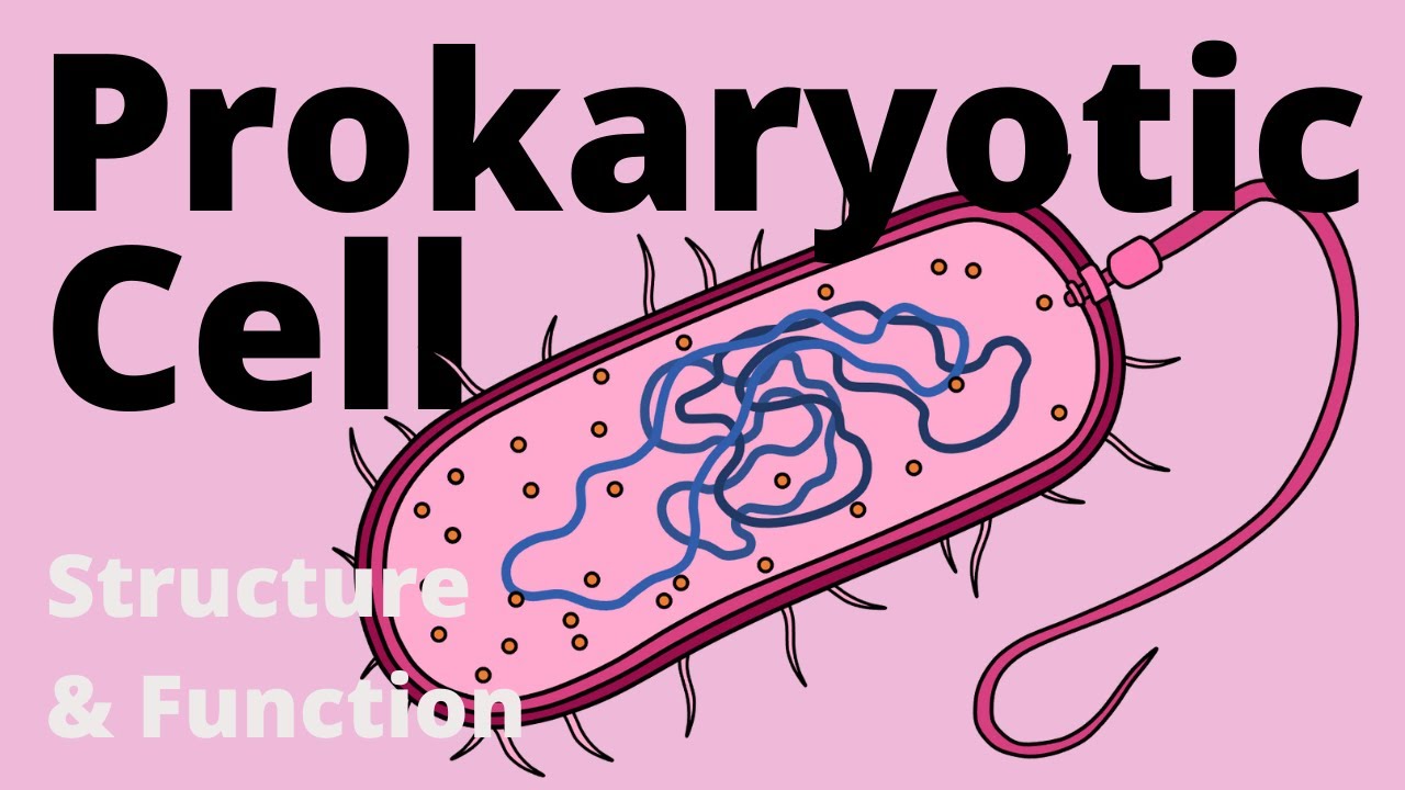
Image taken from the YouTube channel Amoeba Sisters , from the video titled Bacteria (Updated) .
Bacteria, often invisible to the naked eye, are among the most abundant and diverse organisms on Earth. They inhabit virtually every environment, from the deepest ocean trenches to the highest mountain peaks, and even thrive within the bodies of plants and animals.
Their ecological roles are equally vast, ranging from nutrient cycling and decomposition to symbiotic relationships that are essential for the survival of other organisms. Understanding these microscopic powerhouses is crucial for comprehending the complexities of life itself.
The Ubiquitous Nature and Global Significance of Bacteria
Bacteria's adaptability has allowed them to colonize almost every imaginable habitat. They are found in soil, water, air, and extreme environments such as hot springs, glaciers, and radioactive waste sites.
This widespread distribution makes them key players in global biogeochemical cycles, influencing the flow of carbon, nitrogen, and other essential elements. Without bacteria, ecosystems would collapse, and life as we know it would cease to exist.
Furthermore, bacteria are integral to many industrial processes, including the production of food, pharmaceuticals, and biofuels. Their metabolic capabilities are harnessed for bioremediation, cleaning up pollutants, and for various biotechnological applications.
Understanding Bacterial Structure: A Gateway to Scientific Advancements
The study of bacterial structure is more than an academic exercise. It’s a critical foundation for advancements across various scientific disciplines.
In medicine, understanding how bacteria are structured is essential for developing effective antibiotics and combating infectious diseases. Identifying unique structural components of pathogenic bacteria allows researchers to design drugs that specifically target and disrupt these components, without harming human cells.
In biotechnology, a deep understanding of bacterial structure enables the manipulation of these organisms for desired purposes. Genetic engineering of bacteria can lead to the production of valuable proteins, enzymes, and other biomolecules. Knowledge of bacterial cell walls and membranes, for instance, is crucial for developing efficient drug delivery systems and creating novel biomaterials.
In other scientific fields, such as environmental science and astrobiology, the study of bacterial structure provides insights into the evolution of life, the adaptation of organisms to extreme environments, and the potential for life on other planets. Examining the structural adaptations of bacteria in extreme environments, such as the thick cell walls of extremophiles, can provide clues about the limits of life and the conditions under which it can thrive.
This article will explore the essential components of bacterial structure, their functions, and variations, providing a comprehensive overview of these microscopic building blocks of life.
Understanding Bacterial Structure: A Gateway to Scientific Advancements
The study of bacterial structure is more than an academic exercise. It’s a critical foundation for advancements across various scientific disciplines.
In medicine, understanding how bacteria are structured is essential for developing effective antibiotics and combating infectious diseases. Identifying unique structural components of pathogenic bacteria allows researchers to design drugs that specifically target and disrupt these components, without harming host cells.
With a basic understanding established, let's delve into the foundational elements that constitute the bacterial cell. These core components, present in almost all bacteria, are essential for life.
Core Components: The Building Blocks of Bacterial Life
Bacteria, despite their diversity, share several essential components. These are fundamental for survival and reproduction. Let's explore the plasma membrane, DNA (nucleoid), and ribosomes.
The Plasma Membrane: Gatekeeper of the Cell
The plasma membrane, or cell membrane, is the outermost boundary of the bacterial cell. It separates the cell's interior from its external environment.
It's a dynamic and selectively permeable barrier, critical for maintaining cellular integrity.
Structure of the Plasma Membrane
The plasma membrane is primarily composed of a phospholipid bilayer. This means it consists of two layers of phospholipid molecules arranged with their hydrophobic (water-repelling) tails facing inward. The hydrophilic (water-attracting) heads face outward.
Embedded within this bilayer are various proteins. These proteins have diverse functions, including transport, signaling, and energy generation.
Functions of the Plasma Membrane
The plasma membrane performs several critical functions:
-
Selective Permeability: It controls which molecules can enter and exit the cell. This ensures the cell maintains the optimal internal environment.
-
Transport: Membrane proteins facilitate the transport of nutrients into the cell and waste products out of the cell. This is essential for metabolism and maintaining cellular homeostasis.
-
Energy Generation: In many bacteria, the plasma membrane plays a crucial role in energy production through processes like oxidative phosphorylation.
DNA (Nucleoid): The Genetic Blueprint
Unlike eukaryotic cells, bacteria do not have a membrane-bound nucleus. Instead, their genetic material is organized within a region called the nucleoid.
Structure of the Bacterial Genome
The bacterial genome typically consists of a single, circular DNA molecule.
This DNA contains all the genetic information necessary for bacterial growth, reproduction, and survival. The DNA is highly compacted and organized within the nucleoid region. This is achieved through interactions with various proteins.
Role of DNA
DNA serves as the blueprint for all cellular processes. It contains the instructions for synthesizing proteins, replicating the bacterial genome, and responding to environmental changes.
DNA replication ensures that genetic information is accurately passed on to daughter cells during cell division.
Ribosomes: The Protein Factories
Ribosomes are essential cellular structures responsible for protein synthesis. They translate the genetic code encoded in messenger RNA (mRNA) into proteins. Proteins carry out various cellular functions.
Structure of Ribosomes
Bacterial ribosomes are composed of two subunits: a small subunit (30S) and a large subunit (50S).
Each subunit contains ribosomal RNA (rRNA) molecules and ribosomal proteins. These components work together to facilitate the translation process.
Function of Ribosomes
During protein synthesis, ribosomes bind to mRNA molecules and move along the mRNA.
As they move, they read the genetic code and assemble amino acids into polypeptide chains. These polypeptide chains fold into functional proteins. Proteins are the workhorses of the cell, carrying out a vast array of functions.
The Bacterial Cell Wall: Shape, Protection, and Classification
Having explored the core components essential to nearly all bacteria, we now turn our attention to a structure that, while not universally present, plays a critical role in the survival and classification of many bacterial species: the cell wall. This complex structure provides shape and protection and serves as a key differentiator between major bacterial groups.
The bacterial cell wall is not merely a passive barrier. It is a dynamic and intricate structure that determines cell shape, protects against osmotic pressure, and plays a crucial role in the interaction between bacteria and their environment. The most striking difference in cell wall architecture lies between Gram-positive and Gram-negative bacteria, a distinction fundamental to bacterial classification and antibiotic development.
The Peptidoglycan Layer: A Unique Structural Scaffold
The cornerstone of the bacterial cell wall is the peptidoglycan layer, also known as murein. This unique polymer is found exclusively in bacteria and is essential for their survival.
Peptidoglycan is a mesh-like structure composed of two alternating sugar molecules: N-acetylglucosamine (NAG) and N-acetylmuramic acid (NAM). These sugars are linked together in long chains. Attached to NAM is a short chain of amino acids. These amino acid chains are cross-linked between different glycan strands, creating a strong, three-dimensional network.
The specific composition and cross-linking pattern of peptidoglycan can vary slightly between different bacterial species. However, the fundamental structure remains the same. This allows it to provide a rigid framework that protects the cell from lysis due to osmotic pressure.
Gram-Positive vs. Gram-Negative Bacteria: A Tale of Two Walls
The Gram stain, developed by Hans Christian Gram, is a differential staining technique that distinguishes bacteria based on their cell wall structure. This simple staining procedure has revolutionized bacteriology, allowing for rapid classification and identification of bacteria.
The key difference between Gram-positive and Gram-negative bacteria lies in the thickness and complexity of their cell walls.
Gram-Positive Bacteria: A Thick Peptidoglycan Shield
Gram-positive bacteria possess a cell wall characterized by a thick layer of peptidoglycan, which can account for up to 90% of the cell wall's dry weight. This thick peptidoglycan layer is tightly cross-linked, forming a robust and resilient barrier.
In addition to peptidoglycan, Gram-positive cell walls often contain teichoic acids and lipoteichoic acids. These acidic polysaccharides are embedded within the peptidoglycan layer and extend to the cell surface. Teichoic acids contribute to the cell wall's negative charge, which plays a role in ion transport and cell adhesion. Lipoteichoic acids are similar to teichoic acids, but they are anchored to the plasma membrane.
Gram-Negative Bacteria: A More Complex Architecture
Gram-negative bacteria have a more complex cell wall structure than their Gram-positive counterparts. Their cell wall consists of a thin layer of peptidoglycan, surrounded by an outer membrane.
The thin peptidoglycan layer, located in the periplasmic space between the plasma membrane and the outer membrane, accounts for only 5-10% of the cell wall's dry weight.
The presence of the outer membrane is the defining feature of Gram-negative bacteria. This membrane acts as an additional barrier, providing increased protection against antibiotics and other harmful substances.
The Outer Membrane (Gram-Negative Bacteria): An Additional Layer of Defense
The outer membrane is a unique structure found only in Gram-negative bacteria. It is a phospholipid bilayer similar to the plasma membrane, but with several key differences.
The outer leaflet of the outer membrane is composed primarily of lipopolysaccharide (LPS), also known as endotoxin. LPS is a complex molecule consisting of three parts:
-
Lipid A: The hydrophobic anchor that embeds LPS in the outer membrane. It is responsible for the toxic effects of LPS.
-
Core oligosaccharide: A short chain of sugars linked to Lipid A.
-
O-antigen: A long, repeating chain of sugars that extends outward from the cell surface. The O-antigen is highly variable between different bacterial species and is used for serotyping.
The outer membrane also contains porins. These are protein channels that allow small molecules to pass through the membrane. Porins are essential for nutrient uptake and waste removal.
The outer membrane provides a significant barrier to the entry of many antibiotics and other harmful substances. This contributes to the increased antibiotic resistance observed in Gram-negative bacteria.
Cell Wall Variations: Exceptions to the Rule
While the peptidoglycan-based cell wall is a common feature of most bacteria, there are exceptions to the rule. Some bacteria lack a cell wall altogether.
Mycoplasma are a genus of bacteria that naturally lack a cell wall. They are the smallest known bacteria and are characterized by their pleomorphic shape. Because they lack a cell wall, Mycoplasma are resistant to many common antibiotics that target peptidoglycan synthesis. To maintain their structural integrity, Mycoplasma often incorporate sterols into their plasma membrane. They obtain these sterols from their environment.
The absence of a cell wall in Mycoplasma highlights the diversity of bacterial adaptations and the importance of understanding these variations for effective treatment and control of bacterial infections.
Surface Appendages: Interacting with the World
While the cell wall provides a crucial barrier and determines fundamental characteristics like Gram status, bacteria are far from passive entities encased in a rigid shell. To actively engage with their surroundings, bacteria employ a variety of surface appendages, specialized structures that extend outward from the cell.
These appendages are essential for a range of functions, including movement towards nutrients or away from harmful substances, and attachment to host cells or other surfaces. Two of the most prominent types of surface appendages are flagella and pili (also known as fimbriae), each with distinct structures and roles in bacterial behavior.
Flagella: Propelling Bacteria Through Their Environment
Flagella are whip-like appendages that enable bacteria to move through liquid environments. This ability to move, known as motility, is crucial for bacteria to reach favorable conditions, such as areas with high nutrient concentrations, and to escape from adverse environments containing toxins or antibiotics.
The Structure of Bacterial Flagella
Unlike eukaryotic flagella, which have a complex internal structure, bacterial flagella are simpler in design. A bacterial flagellum consists of three main parts:
-
The Filament: This is the long, helical, whip-like structure that extends from the cell surface. It is composed of a protein called flagellin.
-
The Hook: This is a curved structure that connects the filament to the basal body. It acts as a flexible joint, allowing the filament to rotate freely.
-
The Basal Body: This is a complex motor embedded in the cell wall and plasma membrane. It consists of several rings and proteins that drive the rotation of the flagellum.
The Mechanism of Bacterial Motility
Bacterial flagella rotate like a propeller, driven by the basal body motor. The motor is powered by the flow of ions (usually protons) across the plasma membrane. The direction of rotation determines the direction of movement.
When the flagella rotate counterclockwise, they form a bundle that propels the bacterium forward in a smooth, linear motion called a "run."
When the flagella rotate clockwise, the bundle comes apart, causing the bacterium to tumble randomly.
This "run and tumble" behavior allows bacteria to move towards attractants (positive chemotaxis) or away from repellents (negative chemotaxis) in a directed manner.
Types of Flagellar Arrangements
The number and arrangement of flagella can vary significantly between different bacterial species. These variations are often used as a taxonomic characteristic. The main types of flagellar arrangements include:
-
Monotrichous: A single flagellum at one pole of the cell.
-
Amphitrichous: A single flagellum at both poles of the cell.
-
Lophotrichous: A tuft of flagella at one or both poles of the cell.
-
Peritrichous: Flagella distributed over the entire surface of the cell.
Pili/Fimbriae: Anchoring and Attaching
Pili, also known as fimbriae, are short, hair-like appendages that are thinner and shorter than flagella. They are primarily involved in adhesion, allowing bacteria to attach to surfaces, including host cells, inert surfaces, and other bacteria.
Structure and Function
Pili are composed of protein subunits called pilin. They extend outward from the cell surface and can bind to specific receptors on target cells or surfaces.
-
Adhesion: The most important function of pili is to mediate the attachment of bacteria to host cells. This is particularly important for pathogenic bacteria, as it allows them to colonize and infect tissues.
For example, Escherichia coli uses pili to attach to the cells lining the urinary tract, causing urinary tract infections.
-
Biofilm Formation: Pili also play a role in the formation of biofilms, which are communities of bacteria attached to a surface and encased in a matrix of extracellular polymeric substances. Biofilms can be highly resistant to antibiotics and disinfectants, making them a significant challenge in healthcare settings.
-
Conjugation: A special type of pilus, called the sex pilus or F pilus, is involved in bacterial conjugation, a process in which bacteria transfer genetic material to each other. This can contribute to the spread of antibiotic resistance genes.
In summary, surface appendages like flagella and pili are vital tools that bacteria use to navigate their environment, colonize surfaces, and interact with other cells. These structures play crucial roles in bacterial survival, pathogenicity, and the formation of complex microbial communities. Understanding the mechanisms by which these appendages function is essential for developing strategies to combat bacterial infections and harness the beneficial properties of bacteria in various applications.
Additional Structures: Specialized Adaptations
Beyond the fundamental components and surface appendages, certain bacteria boast additional structures that provide enhanced survival capabilities and often contribute to their virulence. These specialized adaptations are not universally present across all bacterial species, but their presence can significantly impact a bacterium's ability to colonize, persist, and cause disease. Among these structures, the capsule stands out as a particularly important determinant of bacterial pathogenicity.
The Capsule: A Protective Shield
The capsule is a layer of viscous, usually gel-like material that surrounds the cell wall of some bacteria. It is typically composed of polysaccharides, although in some species it can be made of other substances, such as polypeptides (e.g., poly-D-glutamic acid in Bacillus anthracis). This structure is external to the cell wall, forming the outermost layer of the bacterial cell.
Composition and Structure
The chemical composition of the capsule varies among different bacterial species, reflecting the diversity of bacterial adaptation strategies. Most bacterial capsules are composed of repeating polysaccharide subunits, forming a thick, hydrated layer. This polysaccharide composition is significant as it is often poorly immunogenic, meaning that the host immune system may not readily recognize and mount a response against it.
The capsule can range in thickness, and some capsules are tightly bound to the cell wall while others are more loosely associated, forming a slime layer. Regardless of its exact structure, the capsule's primary function is to provide a protective barrier between the bacterium and its environment.
Functions and Significance
The capsule plays a crucial role in bacterial survival and virulence through several key mechanisms. One of the most important is its ability to protect bacteria from phagocytosis.
Phagocytosis is the process by which immune cells, such as macrophages and neutrophils, engulf and destroy pathogens. The capsule hinders this process by making it more difficult for phagocytic cells to adhere to and internalize the bacterium. The slippery, often negatively charged surface of the capsule repels the phagocyte, preventing effective engulfment.
Beyond evading the immune system, the capsule also contributes to bacterial virulence in other ways:
-
Adherence: In some cases, the capsule can mediate adherence to host cells or surfaces, facilitating colonization and biofilm formation. This is particularly important for bacteria that cause infections in specific tissues or organs.
-
Protection from Desiccation: The hydrated nature of the capsule helps protect bacteria from dehydration, enabling them to survive in dry or harsh environments. This is crucial for bacterial transmission and persistence.
-
Resistance to Antimicrobial Agents: The capsule can also provide a barrier against certain antimicrobial agents, reducing their effectiveness and contributing to antibiotic resistance.
Clinical Relevance
The presence or absence of a capsule can significantly impact the clinical outcome of bacterial infections. Encapsulated bacteria are often more virulent and more likely to cause severe disease than non-encapsulated strains.
-
Streptococcus pneumoniae, for example, is a leading cause of pneumonia, meningitis, and bacteremia, and its capsule is a major virulence factor.
-
Similarly, Haemophilus influenzae type b (Hib) was a major cause of childhood meningitis before the introduction of Hib vaccines, which target the capsule polysaccharide.
The capsule, therefore, represents a critical target for vaccine development and antimicrobial strategies. Understanding the structure and function of bacterial capsules is essential for developing new ways to prevent and treat bacterial infections.
Video: Bacteria Structure: Unlocking Life's Tiny Secrets
Bacteria Structure: FAQs
Bacteria are incredibly diverse, and their structure plays a crucial role in their survival. Here are some common questions about bacteria structure:
What are the main components of a bacteria structure?
A typical bacteria structure includes a cell wall, cell membrane, cytoplasm containing the genetic material (DNA), ribosomes, and sometimes additional structures like capsules, flagella, or pili. These structures work together to enable the bacteria to function and reproduce.
How does the cell wall contribute to bacteria structure?
The cell wall provides structural support and protection to the bacteria. It maintains the bacteria's shape and prevents it from bursting due to internal pressure. Different types of bacteria have different cell wall compositions affecting how they interact with their environment.
What is the role of flagella in bacteria structure?
Flagella are whip-like appendages that enable bacteria to move. The number and arrangement of flagella vary depending on the type of bacteria, impacting its motility and ability to navigate its surroundings. They are key components of bacteria structure and function.
Is a capsule a common structure in all bacteria?
No, not all bacteria possess a capsule. A capsule is an outer layer made of polysaccharides that surrounds the cell wall in some bacteria. It contributes to the bacteria structure by providing additional protection against phagocytosis and aiding in adherence to surfaces.
So, there you have it – a glimpse into the fascinating world of bacteria structure! Hopefully, you now have a better understanding of these tiny but mighty organisms and their complex construction.